
Novel Non-invasive Imaging Technique May Help Detect Lung Changes Earlier in COPD Patients, Study Says


A relatively new non-invasive imaging technique, called parametric response mapping (PRM), can identify damage in the smallest airways, opening the possibility of detecting specific lung changes earlier (when it is most treatable) in patients with chronic obstructive pulmonary disease (COPD), and allowing doctors to more accurately measure the response to medicines, according to new research.
The results were described in the study, “Non-Invasive Imaging Biomarker Identifies Small Airway Damage in Severe COPD,” and published in the American Journal of Respiratory and Critical Care Medicine.
COPD is characterized by breathing difficulties, and the two most common types of the disease are chronic bronchitis and emphysema. Chronic bronchitis is characterized by inflammation, excessive mucus production, and narrowing of the bronchial tubes, which blocks airflow. Emphysema occurs when the lining of the air sacs is damaged, reducing their elasticity and the ability of the lungs to recoil, resulting in trapped air and the inability to exhale it completely.
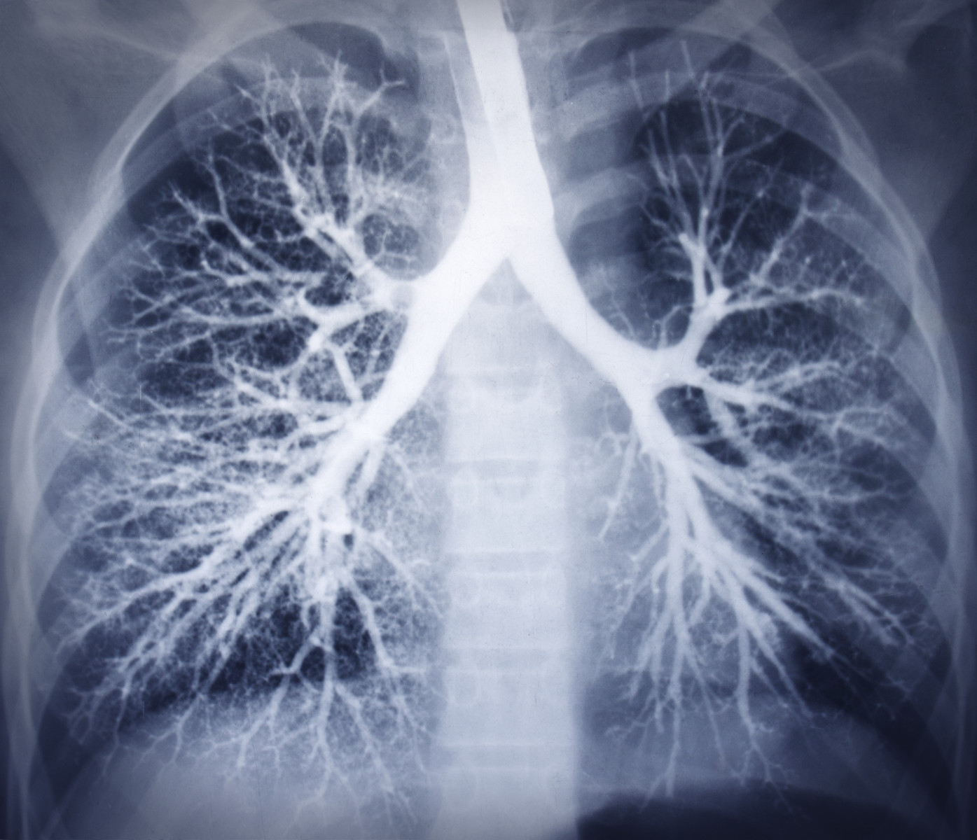
Although computed tomography (CT) imaging can identify emphysema, it has been difficult for physicians to identify abnormalities of the small airways in a non-invasive way. The tiny terminal bronchioles (airways measuring less than two mm in internal diameter) are first damaged in COPD, but they are too small to be visualized on CT imaging, and pulmonary function tests such as spirometry cannot detect these early abnormalities.
Now, an international team led by researchers at Michigan Medicine and funded by the National Heart Lung and Blood Institute (NHLBI) has shown that small airway damage can be identified in COPD patients using PRM, a non-invasive imaging biomarker.
PRM is a sophisticated analysis of computed tomography scans that measures lung density during inhalation and exhalation, processes the resulting images, and classifies each point in three-dimensional space as normal lung parenchyma, functional small airway disease, or emphysema. The result is a color-coded image of the lungs.
The study, which required the collaboration of large, multi-disciplinary teams of radiologists, pulmonologists, thoracic surgeons, and pathologists, involved CT analysis of resected lungs from both healthy controls and patients with severe COPD undergoing lung transplant. Researchers correlated the data with CT scans taken before surgery.
Results showed that PRM could, in a non-invasive way, identify small airway loss, narrowing, and obstruction.
“This is first confirmation that an imaging biomarker can identify terminal bronchial pathology in established COPD, and provides a non-invasive imaging methodology to identify small airway damage in COPD,” the researchers wrote.
“Now we have confidence in our ability to identify airway disease when imaging COPD patients,” MeiLan Han, MD, the study’s senior author, lung specialist, and professor of internal medicine at the University of Michigan, said in the press release.
The patients analyzed in the study had severe disease, therefore the researchers “still need to validate the type of airway disease the PRM technique identifies in patients with milder disease,” Han said. “That type of lung tissue is more difficult to obtain, but we are working on techniques that would allow us to use smaller amounts of lung tissue to make such studies feasible.”
In another NHLBI-funded study, COPDGene, the PRM-defined small airway abnormalities were detected on CT scans of patients with milder disease, raising the possibility of being able to predict loss of lung function in the future.
“These results illustrate the importance of developing non-invasive techniques for improving diagnostic capabilities and advancing new therapies needed to tackle this devastating disease,” said James Kiley, PhD, director of the NHLBI’s lung diseases division. “The refinement of this and similar approaches could also advance the study of COPD at its earliest stages of development.”