Physicians Are First in U.S. to Perform Robotic-assisted Lung Surgery
Written by |
Physicians at Illinois’ Northwestern Memorial Hospital are the first in the United States to successfully perform a robotic-assisted lung volume reduction surgery (LVRS) to extract diseased tissue from the lungs of patients with severe emphysema, a progressive form of chronic obstructive pulmonary disease (COPD). The first procedure was done in late 2018.
The surgery was performed using the da Vinci Xi Surgical System, which allows a surgeon to more accurately remove the diseased parts of the lung. The use of this robotic-assisted procedure over the traditional one can lead to a reduction in pain and scarring, a reduction in the risk of infection, and a shorter recovery time.
“Robotic-assisted technology is revolutionizing the way patients are treated for chronic lung diseases,” Ankit Bharat, MD, surgical director of the Lung Transplant Program at Northwestern Memorial, said in a press release.
Emphysema results from damage to the alveoli (small air sacs in the lung); this damage causes the air in alveoli to become trapped, leading them to expand and rupture.
“By providing this innovative surgical option to our patients, we are changing the paradigm of emphysema management by expanding the possibilities to our patients who desperately need an option to improve breathing to enhance their quality of life and improve overall health,” Bharat said.
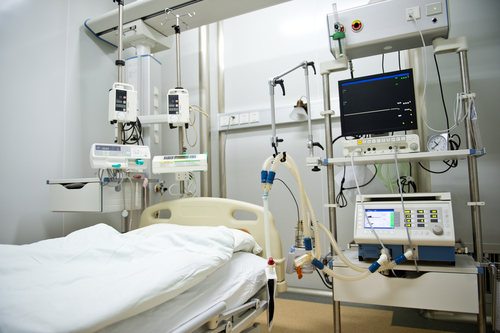
Throughout the robotic-assisted LVRS, the surgeon — who undergoes training in robotic surgery — uses a computer console and looks through a stereoscopic, high-definition monitor to see inside the patient. This allows the surgeon to have a better, more accurate 3D view of the area under surgery.
The apparatus is composed of a tower, which is placed directly on top of the patient during the operation. The tower contains four robot arms, which the surgeon controls and directly replicate the movements of the surgeon. Three of the four arms are used for holding surgical instruments; the fourth arm is used to hold the 3D cameras.
The surgeon, through the use of master controls, moves the arms of the robot in order to create three small incisions on the right side of the chest. These incisions allow the robot to access the lungs and remove the diseased areas, referred to as blebs.
By removing blebs, the remaining lung tissue can function better for optimal breathing. Components of the respiratory system, such as the diaphragm, chest wall, and ribcage, are able to resume a more normal shape and work more efficiently.
As the robotic LVRS only makes three 8-millimeter incisions, as opposed to the larger chest incisions used in traditional surgery, the chances of scarring and infection are much lower. The technique also can shorten the length of stay at the hospital.
“We are excited to expand the therapeutic options we can now offer to a broader range of patients who need surgical intervention and are not responding to medications or pulmonary rehabilitation,” said Ravi Kalhan, MD, pulmonologist and medical director of the LVRS program at Northwestern Memorial.
Kalhan noted that not every patient is a suitable candidate for robotic LVRS, as there are certain criteria. For instance, to be eligible, “patients must be diagnosed with severe COPD with limited exercise capacity after completing pulmonary rehabilitation,” Kalhan said.