COPD’s pulmonary rehab benefits likely tied to oral bacteria changes
12-week program prevented declines in anti-inflammatory Rothia bacteria
Written by |

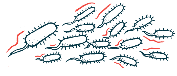
The benefits of pulmonary rehabilitation in people with chronic obstructive pulmonary disease (COPD) may be tied to changes in oral microbiota, the natural community of microbes in the mouth, a study suggests.
Pulmonary rehabilitation for 12 weeks prevented a drop in Rothia bacteria, known for their anti-inflammatory properties, and an increase in species known for pro-inflammatory activity, namely Streptococcus and Lautropia.
The findings were reported in “Responsiveness to pulmonary rehabilitation in COPD is associated with changes in microbiota,” which was published in Respiratory Research.
Pulmonary rehabilitation is a cost-effective approach to managing COPD. It’s a nonpharmacological therapy that incorporates exercise, nutrition, education, and psychological guidance. It’s reported to be three to five times more effective than pharmacological treatments at reducing shortness of breath (dyspnea), improving exercise capacity, and quality of life. Not all patients respond equally to rehabilitation and the reasons for it are poorly understood, however.
COPD severity has been linked to airway microbiota, the community of microbes in the airways that includes oral microbiota. Until now, the impact of pulmonary rehabilitation on airway microbiota hasn’t been known, mainly due to the invasive nature of the procedures to collect samples.
Since microbes in the mouth are associated with those in the airways, saliva represents a “viable noninvasive alternative to sampling the airway microbiota,” said the researchers from Portugal, who compared changes in oral microbiota and inflammation markers in people with COPD who completed a 12-week pulmonary rehabilitation program and those who didn’t, who acted as a control group.
Measuring changes in oral microbiota
The researchers collected saliva samples to analyze the microorganisms in the mouth over five months.
In total, 76 people with COPD participated, including 38 who completed the rehabilitation (intervention group) and 38 who didn’t (control group).
Oral microbiota composition was similar in both groups, comprising mainly bacteria belonging to two phyla: Firmicutes (about 43%) and Bacteroidetes (about 24%).
The dynamics of microbiota composition were markedly different between patients in the two groups, however. After one month of rehabilitation, there were significant changes between both groups in the abundance of four main bacterial species: Veillonella, Prevotella melaninogenica, Haemophilus parainfluenzae, and Streptococcus.
Since changes are likely related to inflammatory responses, researchers measured the levels of 13 cytokines — molecules that mediate and regulate immune and inflammatory responses — at different times in both groups.
Within a month, those who had pulmonary rehabilitation showed significant increases in the levels of two cytokines — interleukin-10 (IL-10) and IL-18 — while no changes were in the control group. Within three months of rehabilitation, IL-10, IL-18, and tumor necrosis factor alpha (TNF-a) levels remained significantly higher in the intervention group.
Rehabilitation led to significant improvements in patient outcomes, including exercise capacity, dyspnea, and disease impact, compared with controls.
In the rehabilitation group, 24% of patients showed reduced dyspnea, 63% had improvements in exercise capacity, as measured by the six-minute walk test (6MWT), and 63% had a lower impact of the disease on their health, as measured by the COPD assessment test (CAT).
A total of 16% of patients responded in all three domains. No domain improvement (nonresponders) was observed in another 16%.
The researchers also compared microbiota composition between responders and nonresponders.
Regarding the response in exercise capacity, responders and nonresponders had significant differences in the frequency of the bacteria Lautropia. Higher numbers were found in the nonresponders by the end of the rehabilitation program.
The levels of inflammatory cytokines were also different between responders and nonresponders. The levels of TNF-a, IL-1beta, IL-18, and IL-10 were higher among nonresponders across all domains, while only TNF-a and IL-10 significantly changed in responders.
For nonresponders, researchers observed Lautropia was positively associated — meaning the greater one, the greater the other — with several pro-inflammatory cytokines. In contrast, the anti‐inflammatory Rothia bacteria were negatively correlated — meaning the greater one, the lower the other — with several pro-inflammatory cytokines in all patients in the responders group. Gemellaceae also was negatively correlated with several inflammatory markers in the responders group.
The researchers also looked for potential bacterial interactions that could be related to patients’ response to rehabilitation. They found that Prevotella bacteria, whose frequency decreased in all nonresponders during treatment, was negatively correlated with Haemophilus, Streptococcus, and Lautropia bacteria for dyspnea, and with Haemophilus for exercise capacity.
Among responders, the frequency of P. melaninogenica increased and was negatively correlated with Kingella for dyspnea, with Lautropia for exercise capacity and disease impact, and with Streptococcus for disease impact. These findings suggest pulmonary rehabilitation is linked to changes in the oral microbiota of COPD patients.
“Specifically, [pulmonary rehabilitation] increases salivary Prevotella melaninogenica and avoids the decline in Rothia and the increase in Streptococcus and Lautropia in responders, which may contribute to the benefits of [pulmonary rehabilitation],” the researchers wrote, noting more research could address the “implications and stability of these findings, clarifying the role of oral microbiota both as a biomarker of [pulmonary rehabilitation] responsiveness and as a therapeutic target.”


